There are two systems used to describe the types of knee dislocation. The Kennedy classification names five directional types — anterior, posterior, lateral, rotatory and medial — by the way the tibia has shifted. The Schenck classification grades the injury from KD I to KD V by which ligaments tore. Both describe a true tibiofemoral dislocation rather than a dislocated kneecap, and most of them are limb-threatening emergencies.
Most people imagine a kneecap that has been pushed to the side when they hear the term “dislocated knee.” That is a different injury. A true dislocation of the knee occurs when the femur and the tibia dislocate at the tibiofemoral joint, and it is that injury the classifications below describe.
An example to help clarify this: If a driver’s knee is flexed during an impact and the dashboard drives the tibia posteriorly, both cruciates tear, and the popliteal artery behind the joint is stretched or torn, what has occurred is a true posterior tibiofemoral dislocation — and the clock on that leg starts immediately.
Is a “dislocated knee” the same as a dislocated kneecap?
No, and separating them is the first thing worth doing. A dislocated kneecap occurs when the kneecap slides out of its trochlear groove, usually due to a hard rotational change in direction. Cleveland Clinic describes it as an injury that is often reduced without sedation and heals over weeks to months, with no threat to the leg’s blood supply.
A true tibiofemoral dislocation is a different category of injury. It follows high-energy trauma, tears multiple ligaments by definition — making it a multiligament knee injury (MLKI) — and can cut off circulation to the lower leg.
For scale: approximately one person per 100,000 experiences a true knee dislocation every year, roughly 1 in 5,000 orthopaedic injuries, according to Wikipedia’s overview of knee dislocation. Every one of the types of knee dislocation described below refers to that injury — a true tibiofemoral dislocation — and not to patellar instability.
What are the five types of knee dislocation by direction?
They are described by Kennedy classification by the position of the tibia relative to the femur:
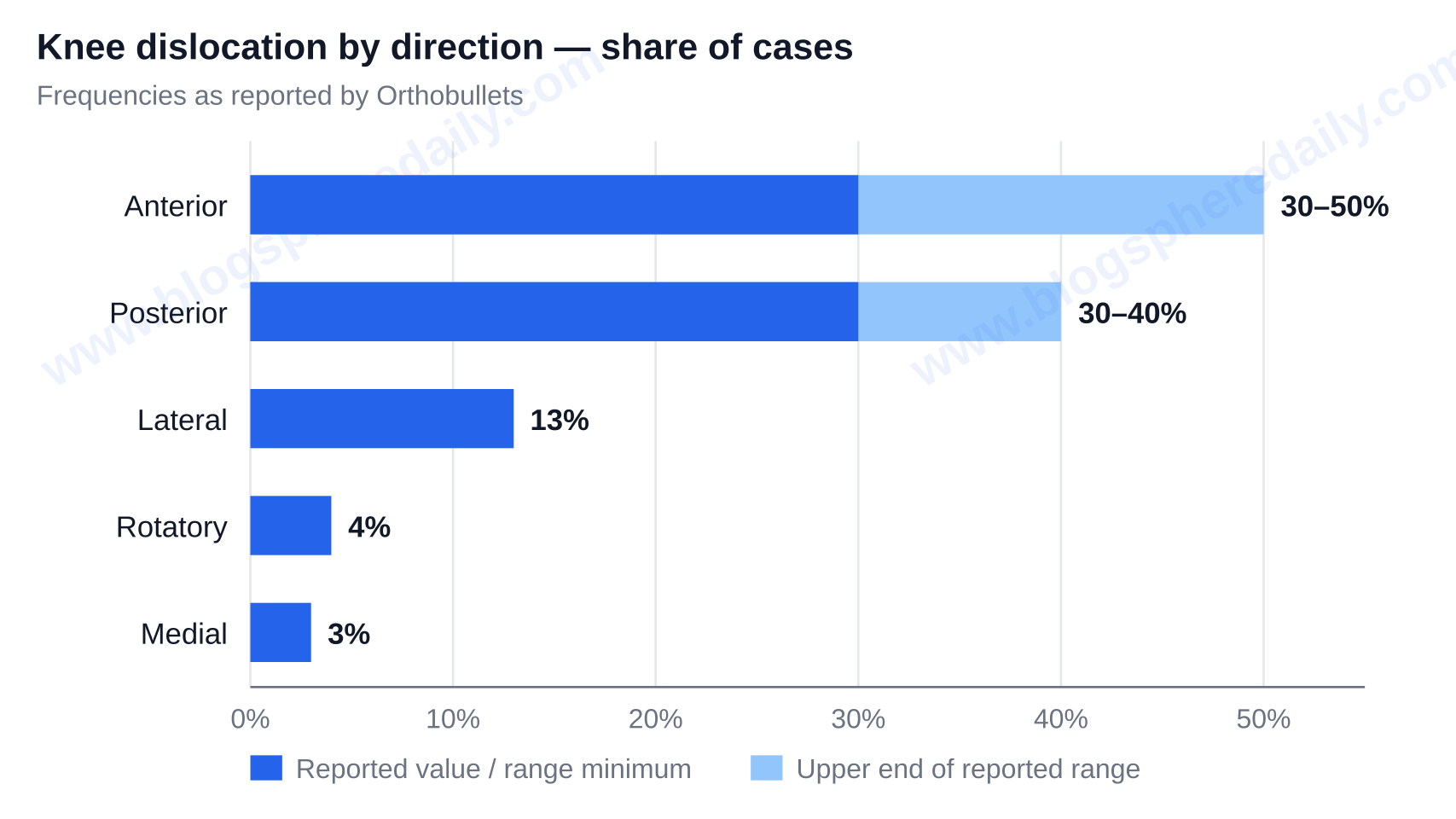
- Anterior (30–50%) — the most common. Hyperextension of the knee usually results in a tear of the posterior cruciate ligament (PCL), and carries the highest rate of common peroneal nerve injury.
- Posterior (30–40%) — an axial load on a knee in a flexed position, the classic “dashboard” mechanism. Highest rate of vascular injury and of complete popliteal artery tear.
- Lateral (13%) — a valgus or varus force, usually results in a tear of both cruciates.
- Rotatory (4%) — a posterolateral dislocation is the most common type of this group and is usually irreducible by closed means.
- Medial (3%) — a tear of the PCL and a disruption of the posterolateral corner is common.
Orthobullets has listed the frequencies of each of these types of knee dislocation. What each direction predicts about risk matters more than the label itself.
Which type of knee dislocation is most dangerous?
The answer to the question, “what type do I have?” is not as useful as the answer to, “what does my type predict?” Each of the figures below can be found throughout different sections of Orthobullets’ knee dislocation page (classification, evaluation and complications). Joining them into a single table tells a different story:
| Direction | Frequency | Mechanism | Ligaments typically torn | Popliteal artery risk | Peroneal nerve risk | Closed reduction |
|---|---|---|---|---|---|---|
| Anterior | 30–50% | Hyperextension | PCL | High (40–50% for anterior/posterior) | Highest of all types | Usually yes |
| Posterior | 30–40% | Axial load, flexed knee (dashboard) | Both cruciates | Highest; most complete arterial tears | Present | Usually yes |
| Lateral | 13% | Valgus/varus force | ACL and PCL | Lower than anterior/posterior | Present | Usually yes |
| Rotatory | 4% | Rotational force (posterolateral most common) | Varies by subtype | Lower than anterior/posterior | Present | Usually no — often irreducible |
| Medial | 3% | Varus force | PCL and posterolateral corner (PLC) | Lower than anterior/posterior | Present | Usually yes |
The baselines that anchor the table: vascular injury occurs in 5–15% of all knee dislocations, but in 40–50% of the anterior and posterior types. Overall, injury to the peroneal nerve occurs in 25% of cases, and Wikipedia reports popliteal artery injury in about 20% of cases with amputation of part of the leg required in just over 10%.
Why the anterior and posterior types dominate the vascular risk
Read the table as a shape, not a list. The anterior type is the most common (and the one most likely to result in foot drop). The posterior type is the one most likely to take the limb, because it tears the artery outright. The rotatory type threatens the reduction itself — a posterolateral type dislocation can not be put back into place without surgery.
What the directional type tells you — and what it doesn’t
Direction predicts risk. It never excludes it. A foot that feels warm and a pulse that feels present do not clear any directional type. An objective vascular measurement is necessary, covered below.
What is the Schenck classification (KD I to KD V)?
This second grading system is based on what ligaments were torn, rather than where the tibia went. The terminology used in charts, operative notes and publications is as follows:
- KD I — multiligament injury with one cruciate ligament intact.
- KD II — both cruciate ligaments torn with both collaterals intact.
- KD III — both cruciate ligaments torn with one corner: KD-III-M (PMC/MCL) or KD-III-L (PLC/LCL).
- KD IV — ACL, PCL, PMC and PLC all torn.
- KD V — multiligament injury with an associated periarticular fracture (fracture-dislocation).
In 1997, Wascher and colleagues modified this system by adding suffixes: -C for arterial injury, -N for nerve injury. KD-III-L-C means both cruciate ligaments, the posterolateral corner, and a damaged artery.
A common confusion worth clearing is that a “Grade 3 knee dislocation” typically means a KD III injury on Schenck’s scale — both cruciate ligaments and a corner torn. This is different from a Grade III ligament sprain, which means a single ligament is completely torn. Different systems of classification, different elements being assessed.
Where the KD classification is changing — and where KD I breaks down
Neither classification is clear cut. A global modified Delphi consensus of 27 experts, published in the Journal of ISAKOS in 2022 (Vol 7, Issue 5, 95–99), agreed on four factors any classification should include: vascular injury (89% agreement), common peroneal nerve injury (78%), number of ligaments torn (78%), and presence of an open injury (70%). The panel endorsed extension of Schenck’s classification with an “open injury” suffix. Open versus closed changes management in a concretely different way: contamination, the need for immediate surgical exploration, and a different reconstructive time-window.
KD I is the weakest part of the classification. A 2023 multicentre study on 227 knee multiligament injuries, published in Orthopaedic Journal of Sports Medicine, labelled 63 (27.8%) of the injuries as KD I on ligament pattern — and only 12 of those 63 (19.0%) had radiologically confirmed tibiofemoral dislocation, representing just 5.3% of the total cohort.
Of those 12 confirmed dislocations, 2 (16.7%) had nerve injuries and 1 (8.3%) had a vascular injury. Cluster risk was seen in specific injury sub-patterns. For example, PCL+MCL (KD I-DPM) produced a popliteal occlusion in 1 of 2 cases, and ACL+LCL (KD I-DAL) a peroneal palsy in its single case. The authors of the study recommend applying Schenck only to bicruciate injuries, or to single-cruciate injuries if dislocation is confirmed on radiographs. A 2025 systematic review in Arthroscopy, Sports Medicine, and Rehabilitation comes to a similar conclusion at the other end of the scale: KD V is under-specified and needs more reporting detail.
Takeaway: KD I is not simply a “mild” dislocation. It is a term that may or may not describe a joint that actually dislocated.
Can a dislocated knee pop back in by itself?
Yes — and that is what makes this injury so easy to underestimate. Orthobullets reports that approximately 50% of dislocated knees reduce (pop back in) on their own before the patient even arrives at the hospital, and therefore are misdiagnosed.
A normal-looking knee and a normal X-ray do not rule out a true tibiofemoral dislocation. The finding that should override a reassuring exam is gross multidirectional instability with a high energy injury. Until the vascular status is cleared, treat this as a reduced dislocation.
The opposite problem is an irreducible dislocation, classically a posterolateral rotatory dislocation. A medial dimple in the skin indicates a button-holed capsule. Closed reduction should stop at this point because the joint needs open reduction. For patients, the framing is simple: the knee going back in relieves the pain, but not the risk.
Is a dislocated knee a medical emergency, and what are the red flags?
Yes, a true tibiofemoral dislocation is managed as a limb-threatening emergency, and therefore in the first few hours, the vascular check matters more than the type.
Two elements are relevant for decision-making. In a prospective study of 38 cases of knee dislocation, all 11 (29%) cases with an ankle-brachial index (ABI) less than 0.90 had arterial injury requiring surgery, while all 27 cases with an ABI of 0.90 or higher had no vascular injury on serial examination or duplex ultrasonography. The second element is the ischaemia clock. The amputation rate approaches 86% once ischaemia time passes roughly six to eight hours, making revascularisation within this time frame the decisive variable.
Red flags that belong in an emergency department include: a pale, cold, or pulseless foot; rapidly expanding calf swelling; inability to lift the foot or numbness across the dorsum (common peroneal nerve palsy, foot drop); obvious deformity; an open wound over the joint; or pain out of proportion after high-energy trauma. The question to ask the clinician is direct — has an ABI been measured, and does this need CT angiography?
How are the types of knee dislocation diagnosed and treated?
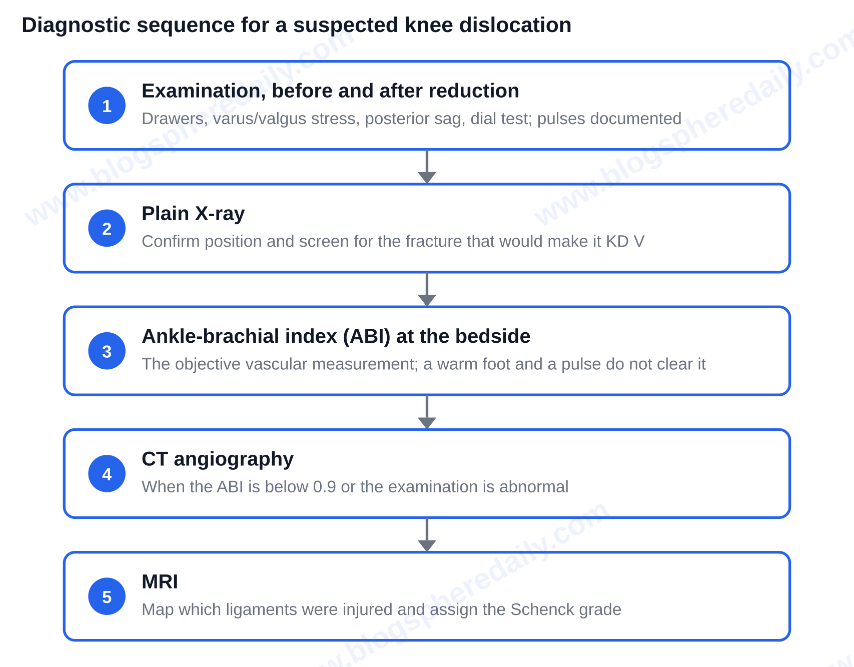
Examination is mapped to specific directions: anterior and posterior drawers, varus and valgus stress tests, the posterior sag test, and the dial test for rotatory patterns. These should be performed before and after reduction, with pulses and distal neurological findings evaluated and documented. Imaging then follows a sequence: plain X-ray to confirm position and screen for the fracture that would make it KD V, ABI at the bedside, CT angiography when the ABI is below 0.9 or the exam is abnormal, and MRI to evaluate which ligaments were injured and to assign the Schenck grade.
Treatment initially includes closed reduction with longitudinal traction under procedural sedation. This is followed by a repeat assessment of the neurovascular status and splinting. When that is not enough, further treatment may include external fixation for a knee that will not stay reduced, open reduction for irreducible posterolateral dislocations, emergency vascular repair or bypass for arterial injury, and fasciotomy where compartment syndrome threatens. Ligament injuries are treated in a staged fashion with the vascular and soft-tissue problems dealt with first, then acute or delayed reconstruction of the cruciates and corners depending on stability, skin condition and fracture pattern.
One shift worth naming in who sustains these injuries. Alongside motor vehicle collisions, contact sport and falls from height, ultra-low-velocity knee dislocations are increasing alongside the rising prevalence of obesity. A 2025 current-concepts review reports vascular injury requiring intervention in about 5.6% of tibiofemoral dislocations in the general population, 7.2% in obese patients and 11.3% in morbidly obese patients, with complication rates increasing 9.2% for each one-unit increase in BMI and smaller ultra-low-velocity series reporting amputation rates of 12–28%.
How long does recovery take, and what should you expect?
Expect days to weeks of immobilisation or external fixation, protective range of motion early to prevent stiffness, ligament reconstruction weeks to months out, and commonly 9–12 months before a return to demanding activity — longer in cases of KD IV and KD V.
There are two major issues in the long term. Orthobullets reports stiffness as the most common complication at 38%, with residual instability affecting 37%. Less likely but defining outcomes include permanent foot drop from peroneal nerve injury, post-traumatic arthritis, and amputation in just over 10% of dislocations overall.
There are many factors which affect prognosis, and they are worth naming plainly: how many ligaments tore, whether the artery or nerve was involved, whether the injury was open, how long the limb was ischaemic, and BMI. In the case of a true knee dislocation the honest expectation is to have a stable, functional limb — not the same knee as before.
The one thing to remember
Both systems for grading the types of knee dislocation exist to predict a single thing: how much danger the limb is in. Anterior is the most common, posterior the most likely to tear the popliteal artery, rotatory the most likely to be irreducible, and KD I the least trustworthy label on the list. In the setting of a high-energy injury, if the knee was left grossly unstable — even if it popped back into place and the X-ray reads normal — get to an emergency department and ask for an ankle-brachial index, because an ABI under 0.90 and the six-to-eight-hour ischaemia window are what decide the outcome. For more like this, explore Blog Sphere Daily.
FAQ
What ligaments are torn in a knee dislocation?
By definition a true tibiofemoral dislocation is a multiligament knee injury, so at least two ligaments are disrupted. The Schenck classification grades patterns of multiligament knee injuries and defines KD I as having one cruciate ligament intact, KD II as both cruciates injured with the collaterals intact, KD III as both cruciates and either the posteromedial corner (KD-III-M) or the posterolateral corner (KD-III-L) injured, KD IV as the ACL, PCL, posteromedial and posterolateral corners all injured, and KD V as a multiligament injury with a periarticular fracture. The direction of the dislocation can also give an indication of the injury pattern. Anterior dislocations usually involve the PCL and medial dislocations usually involve the PCL as well as the posterolateral corner.
Which knee dislocation is hardest to put back in place?
According to Orthobullets, approximately 4% of knee dislocations are rotatory. The most common type of rotatory dislocation is the posterolateral type, which are usually irreducible by closed means. Closed reduction of knee dislocations should stop if there is a skin dimple on the medial side of the knee. This is the classic indication that the joint capsule has buttonholed, and at that point the knee needs open reduction in theatre.
Can you walk on a knee that has been dislocated and reduced?
Not safely, and the patient must be assessed first. One of the problems with knee dislocations is that roughly 50% self-reduce before the patient gets to the hospital and they are often misdiagnosed. A knee that feels like it can be walked on could still be a reduced dislocation with a damaged nerve or blood vessel behind it. Early management is immobilisation or external fixation with protected range of motion, and a return to demanding activity commonly takes 9–12 months — longer for KD IV and KD V injuries.
Does a knee dislocation always show up on an X-ray?
No. Because about half of these injuries reduce spontaneously before the patient is seen, a plain X-ray done afterward can be completely normal. Therefore, a knee that shows gross multidirectional instability after a high-energy injury should always be considered a reduced dislocation until the vascular status is cleared — with a bedside ankle-brachial index (ABI) first, a CT angiogram if the ABI falls below 0.9 or the exam is abnormal, and an MRI to map the torn ligaments.
What does the -C or -N after a KD grade mean on my imaging report?
Those suffixes are from Wascher’s 1997 modification of Schenck’s classification. A -C indicates the presence of an arterial injury, and a -N indicates the presence of a nerve injury. So KD-III-L-C describes both cruciates torn plus the posterolateral corner, with arterial involvement. The 2022 Delphi consensus published in the Journal of ISAKOS recommended adding a suffix for open injuries, since the expert panel agreed that open wounds, vascular injury, peroneal nerve injury and the number of ligaments torn are the four factors a classification needs to capture.